TMI D-Angle
D-Angle
The validation objects used to investigate reconstruction accuracy as a function of the orientation of the endoscope are shown in Fig. 1 and comprise five parts of a liver (flat, convex, with discontinuity, with a hole, with fatty tissue), each reconstructed for two different distances (5.5 cm and 7 cm) and two orientations of the endoscopes: Angles of 90° and 60° measured between the view direction of the endoscope and the tangent plane. This yielded a total of n = 20 surfaces (10 for each angle). Details can be found in [1].
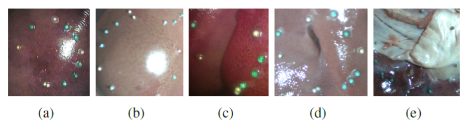
Figure 1: Different shapes of the liver: flat (a), convex (b), with discontinuity (c), with hole (d), with fat (e). The circles represent the region of interest, and the markers were used for registration with computed tomography (CT) data (cf. Fig. 2).
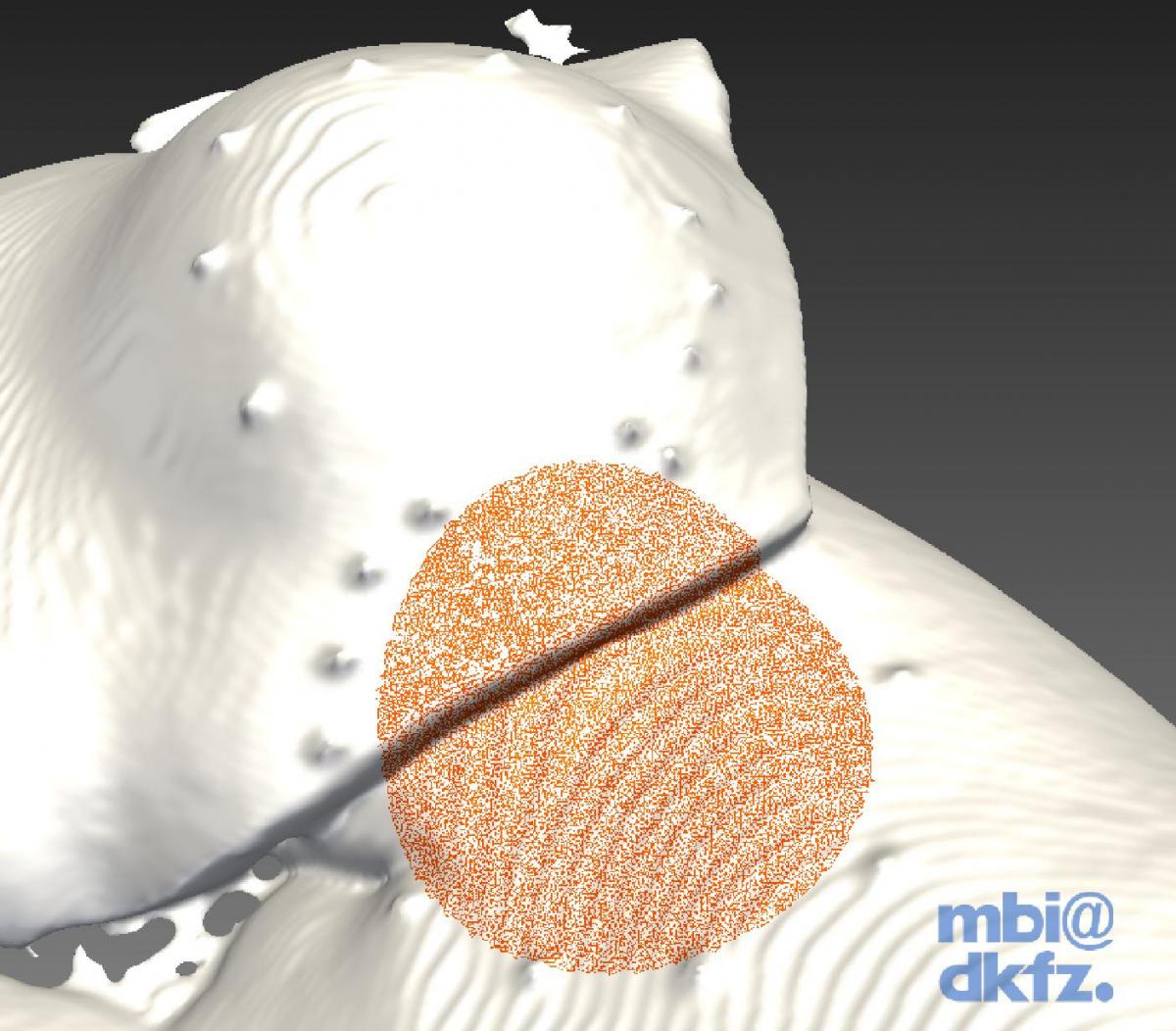
Figure 2: Extraction of a sphere-shaped region (red) from the reference computed tomography (CT) surface.
Download all data sets
Download validation tool
References:
[1] Maier-Hein L, Groch A, Bartoli A, Bodenstedt S, Boissonnat G, Chang PL, Clancy NT, Elson DS, Haase S, Heim E, Hornegger J, Jannin P, Kenngott H, Kilgus T, Müller-Stich B, Oladokun D, Röhl S, Dos Santos TR, Schlemmer HP, Seitel A, Speidel S, Wagner M, Stoyanov D. Comparative Validation of Single-shot Optical Techniques for Laparoscopic 3D Surface Reconstruction. IEEE T Med Imag (in press), 2014