Thyroid Segmentation in Ultrasonography Dataset
The reliable and accurate segmentation of the thyroid in ultrasonography is an open challenge. The segmentation in 3D data can be used to compute the volume, which is an indicator of pathological changes in the thyroid.
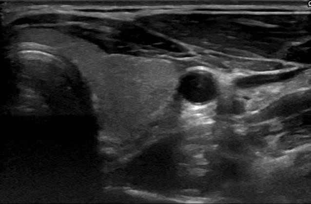
For the development of segmentation techniques reconstructed volumes from tracked ultrasound sweeps are provided below, paired with the matching ground truth. The recordings were performed with a GE Logiq E9 XDclear 2.0.
The acquisition of the data as well as the manual ground truth segmentation with liveWire were performed by a medical expert. The eight DICOM files each contain one lobe of the thyroid. All of the scans contain healthy thyroids. The area per slice that contains the thyroid region has a resolution of around 240x120 px.
The dataset can be downloaded here


If you use this dataset, please cite the following paper:
T. Wunderling, B. Golla, P. Poudel, C. Arens, M. Friebe and C. Hansen, Comparison of thyroid segmentation techniques for 3D ultrasound. Proceedings of SPIE Medical Imaging, Orlando, USA, 2017.