TMI D-Distance
D-Distance
The objects used to investigate reconstruction accuracy as a function of distance are shown in Fig. 1 and comprise five parts of a liver (flat, convex, with discontinuity, with a hole, with fatty tissue), two parts of a kidney (homogeneous, with cut), two parts of a porcine heart (little texture, high texture), and fatty tissue, each acquired with a direct view of the endoscopes on the objects and reconstructed for two different distances: 4 cm, and 7 cm. This yielded a total of n = 20 surfaces (10 for each distance). Details can be found in [1].
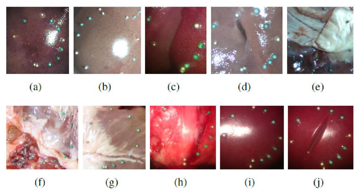
Figure 1: (a)-(e): Different shapes of the liver: flat (a), convex (b), with discontinuity (c), with hole (d), with fat (e). (f)-(j): Different shapes of the heart (f/g), fatty tissue (h), and a kidney without (i) and with (j) cut. The circles represent the region of interest, and the markers were used for registration with computed tomography (CT) data (cf. Fig. 2).
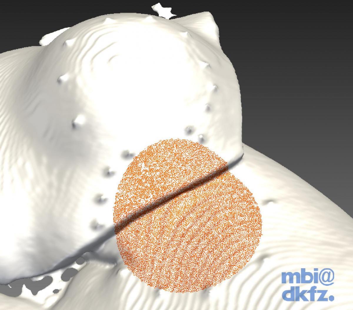
Figure 2: Extraction of a sphere-shaped region (red) from the reference computed tomography (CT) surface.
Download all data sets
Download validation tool
References:
[1] Maier-Hein L, Groch A, Bartoli A, Bodenstedt S, Boissonnat G, Chang PL, Clancy NT, Elson DS, Haase S, Heim E, Hornegger J, Jannin P, Kenngott H, Kilgus T, Müller-Stich B, Oladokun D, Röhl S, Dos Santos TR, Schlemmer HP, Seitel A, Speidel S, Wagner M, Stoyanov D. Comparative Validation of Single-shot Optical Techniques for Laparoscopic 3D Surface Reconstruction. IEEE T Med Imag (in press), 2014